
Photo Credit: Kellen Bakovich/CVMBS
Revolutionizing Equine Diagnostics: The Latest in CT Technology
Discover how advanced CT technology is transforming equine diagnostics, improving precision, and creating new possibilities in veterinary care.
The introduction of computed tomography (CT) has forever changed the landscape of diagnostics. First developed for human healthcare in the 1970s, CT quickly gained recognition for its ability to produce detailed anatomical images. By the 1980s, it found its way into veterinary medicine, and its impact in equine care has grown ever since. Today, CT stands as a cornerstone of equine diagnostics, driven by innovations that are broadening its accessibility and expanding its applications.
What Is Computed Tomography (CT)?
CT technology takes imaging a step beyond traditional X-rays by creating detailed, three-dimensional views of a horse’s anatomy. Unlike standard X-rays, which produce flat, overlapping images, CT allows veterinarians to examine cross-sectional “slices” of bones, tissues, and organs. This enables a level of precision that is crucial for diagnosing complex conditions.
CT scans are completed by combining multiple X-ray images taken from various angles. The result is a highly detailed model of the scanned area, allowing veterinarians to detect subtle issues, track injury progression, and plan surgical procedures with confidence.
Compared to other imaging modalities like MRI, CT offers faster imaging, making it particularly useful for capturing detailed images of standing horses. It also excels at visualizing bone and specific soft tissue structures, filling diagnostic gaps where other methods may fall short.
Types of CT Scanners in Equine Medicine
CT scanners used in equine care fall into two primary categories, each designed for specific needs:
• Fan-beam scanners: These systems are known for producing exceptionally detailed images, especially for soft tissue and intricate structures. However, they often require the horse to be under general anesthesia to pass through the enclosed, doughnut-shaped scanner—particularly for areas like the neck or upper limbs.
• Cone-beam scanners: With an open design, cone-beam scanners are ideal for imaging the lower limbs of standing horses. These scanners capture larger areas in a single rotation, offering a safer and more practical solution for certain diagnostics without requiring anesthesia.
Recent advancements have also introduced systems that combine the high-resolution imaging capabilities of fan-beam scanners with the accessibility of standing, open designs. This evolution has made CT an even more valuable tool in equine veterinary care.
How CT Is Used in Equine Medicine
CT has become an indispensable diagnostic tool for a variety of equine conditions. Some of its most impactful applications include:
• Lameness Evaluation: CT scans provide unparalleled detail for detecting bone issues like fractures, arthritis, and subtle stress injuries that might be missed on traditional X-rays. These insights are key for understanding and addressing lameness.
• Dental and Sinus Health: The complex anatomy of a horse’s head can make diagnosing dental infections, sinusitis, and nasal tumors challenging. CT simplifies this process, offering highly detailed images that aid in accurate diagnosis and treatment planning.
• Neck and Spine Imaging: Neurologic conditions, such as Wobbler syndrome, and spinal issues like arthritis or compression can be diagnosed with greater precision using CT scans. These detailed images are critical for treating horses experiencing stiffness or neurological symptoms.
•Surgical Planning: For complex procedures like fracture repair, CT provides surgeons with an accurate roadmap, improving surgical outcomes.
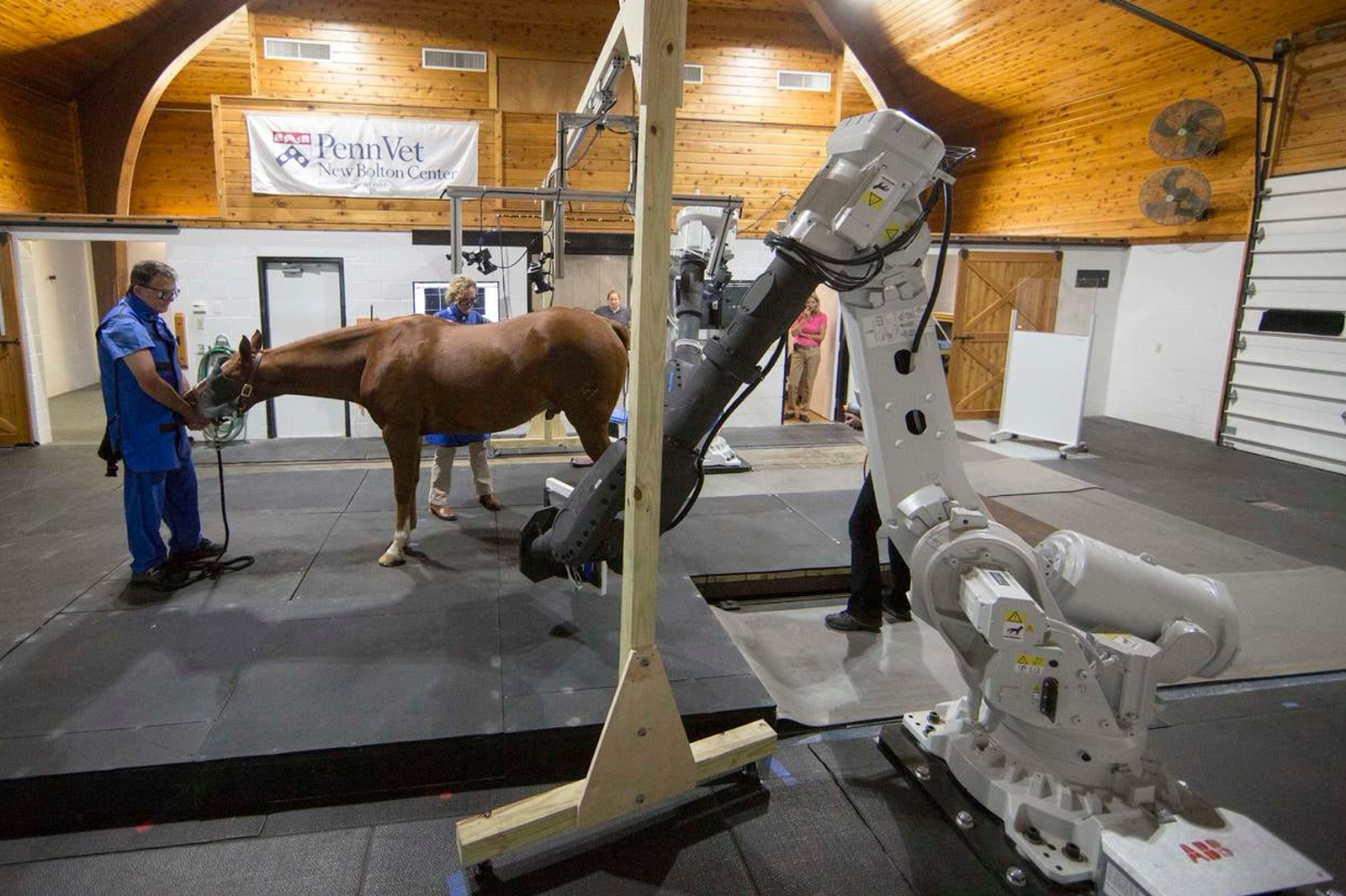
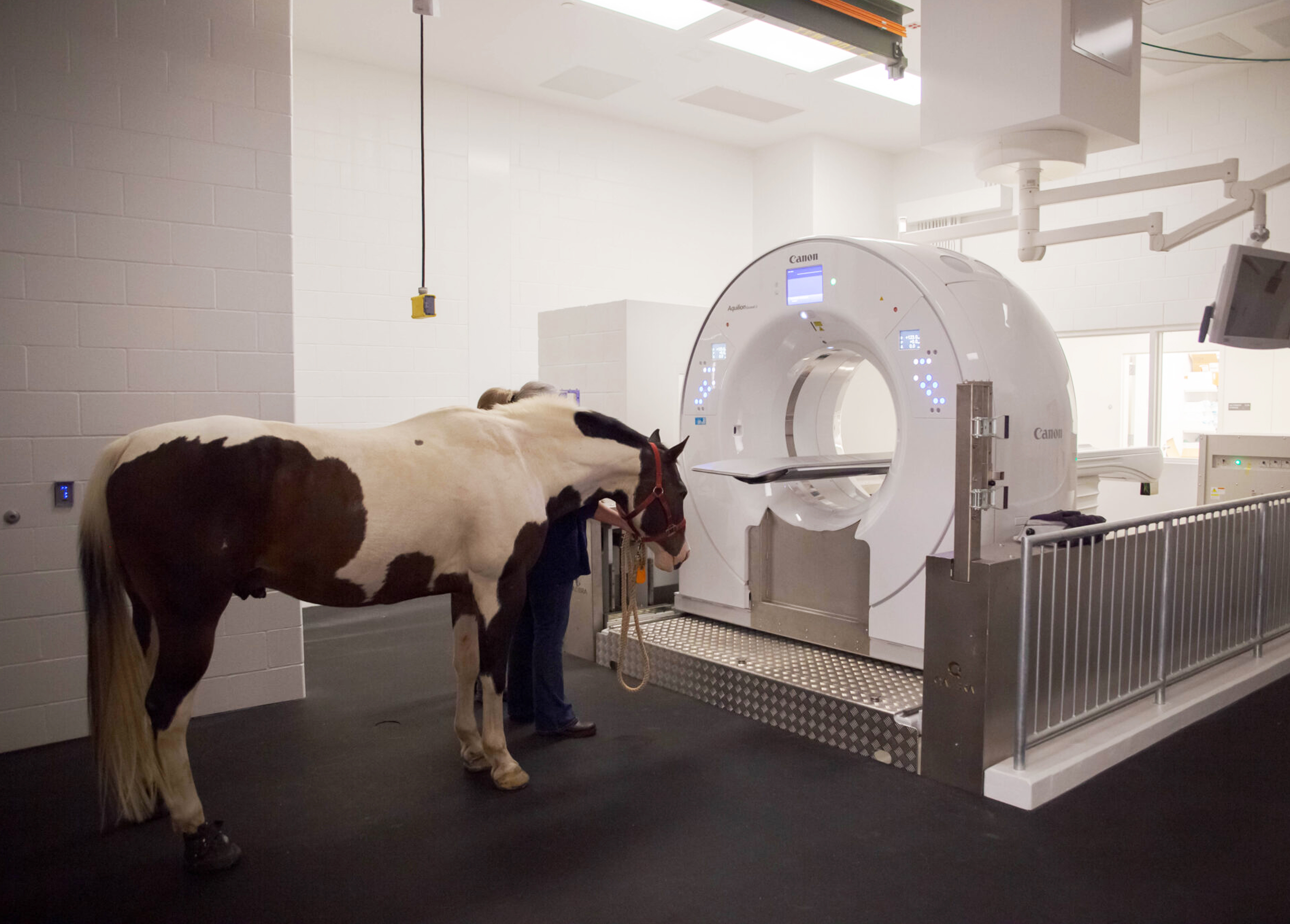
University of Pennsylvania’s New Bolton Center Hospital for Large Animals
Advancements in Standing CT Technology
One of the most significant advancements in recent years is the development of standing CT systems. These systems allow veterinarians to image horses in natural, weight-bearing positions, eliminating the need for general anesthesia.
Standing CT has become a go-to solution for imaging distal limbs, necks, and even the pelvis. For high-performance horses like racehorses, this technology is invaluable for early detection of bone stress or injuries, allowing for preventive care before minor issues escalate into major problems.
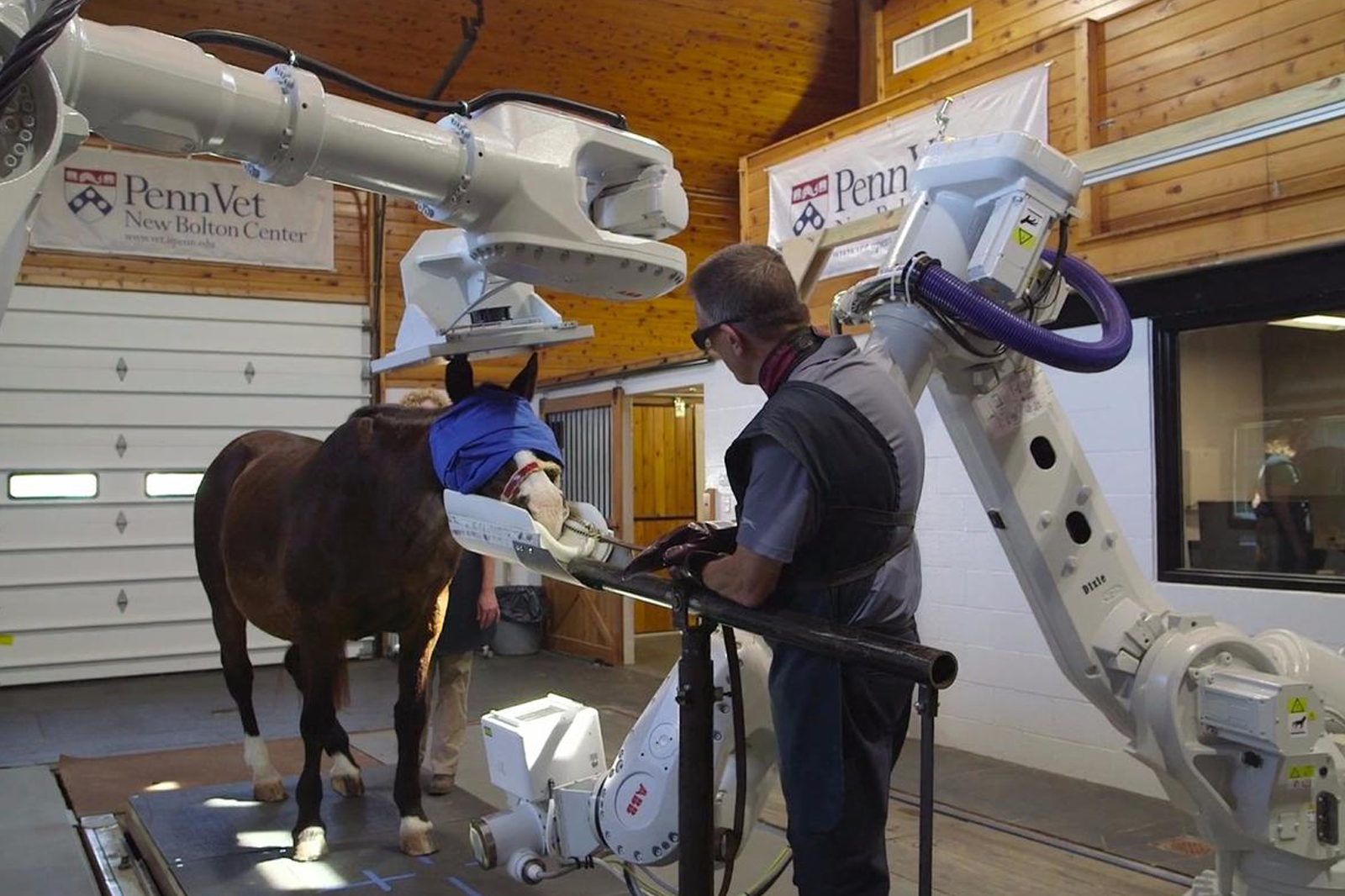
University of Pennsylvania’s New Bolton Center Hospital for Large Animals
Combining CT with PET Imaging
A groundbreaking innovation in equine diagnostics is the combination of CT with positron emission tomography (PET). While CT provides detailed anatomical views, PET highlights areas of increased metabolic activity—such as inflammation or active bone remodeling—using specialized tracers.
When combined, these technologies offer an unparalleled level of precision. Vets can not only pinpoint problem areas but also assess the activity within those areas, enabling more effective diagnosis and monitoring of injuries.
CT Scans for Orthopedic Issues
CT scans have become a vital tool for identifying and treating lameness in horses, thanks to their ability to catch subtle bone problems like stress fractures and bone bruises. These insights help vets pinpoint issues and create targeted treatment plans.
The introduction of standing CT technology has taken this even further. Today’s systems can scan areas like the pelvis, back, and upper limbs without requiring anesthesia. This has been a game changer for racehorses and other high-performance horses, allowing early detection of bone stress and injuries before they become severe.
Transforming Equine Diagnostics
As advancements like standing CT and PET imaging continue to reshape equine medicine, they offer unparalleled opportunities for earlier detection, precise treatment, and improved outcomes for horses. At Kodiak Collective EQ, we’re passionate about sharing the latest in equine health technology and innovation, empowering equestrian professionals to stay ahead of the curve.